


Introduction
Materials and Methods
Sample Collection and Virus Identification
Host Reaction Experiments
Sequence Analysis and Phylogenetic Analysis
Results and Discussion
Introduction
Gomphrena globosa is an annual herbaceous flower belonging to the Amaranthaceae family. It is a horticultural crop widely cultivated for ornamental and cut-flower purposes. Research on plant viruses that cause natural infections in Gomphrena globosa is limited, with only two species (potato virus X and tomato bushy stunt virus) having been identified (Appiano et al., 1978; Pennazio and Redolfi, 1980).
Cucumber mosaic virus (CMV) is a representative plant virus that infects over 1,200 plant species globally. It causes considerable agricultural damage by inducing different symptoms such as mosaic patterns, yellowing, and deformities (Palukaitis et al., 1992). CMV is broadly classified into subgroups I and II based on the genetic characteristics, with subgroup I further subdivided into IA and IB. CMV isolates exhibit different biological characteristics, such as geographic distribution and symptom expression in hosts, depending on their genetic diversity (Mochizuki and Ohki, 2012; Roossinck et al., 1999). The CMV genome consists of three single-stranded RNA molecules, of which RNA3 encodes the movement protein (MP) and the coat protein (CP). Virus-host interactions determine the differences in the host response and disease symptoms among the CMV isolates, and different factors within the viral genome are involved in this process. In particular, MP and CP, encoded by RNA3, are involved in intercellular movement and particle formation and are major determinants of host range and disease symptom expression (Choi et al., 2004; Ryu et al., 1998). Thus, comparative analysis of the biological and molecular characteristics of CMV isolates derived from specific host plants can improve understanding of viral pathogenicity, host adaptability, and genetic diversity.
Gomphrena globosa is widely employed as an experimental host for assessing the pathogenicity and host responses of different CMV isolates. However, studies on the biological and molecular characteristics of CMV isolated from wild Gomphrena globosa are absent. Meanwhile, although the CP sequence (KP455738.1) of CMV-Go5, derived from Iranian G. globosa, is registered in GenBank, the biological characteristics of this isolate, including its host response, have not been reported. Thus, elucidating the biological and molecular characteristics of CMV naturally infecting G. globosa could provide crucial foundational data for understanding CMV host adaptability and diversity among isolates. Accordingly, this study examined the biological and molecular characteristics of two CMV isolates (CMV-GomCC1 and CMV-GomCC2) isolated from G. globosa collected from two different cultivation sites in Chuncheon, Gangwon Province, South Korea, and sought to elucidate the characteristics of CMV isolates derived from G. globosa via phylogenetic and sequence comparisons with the existing CMV isolates.
Materials and Methods
Sample Collection and Virus Identification
In 2024, G. globosa samples with mosaic symptoms were collected from two different locations in Chuncheon, Gangwon Province, South Korea (Fig. 1). Total RNA was extracted from the symptomatic samples using the BCSTM Plant RNA Prep Kit (Biocube System Inc., Suwon, Korea), and cDNA was synthesized using M-MLV reverse transcriptase (Promega, Madison, WI, USA). The 3’-end reverse primers from each virus detection primer set were used for the reverse transcription reaction. Subsequently, a polymerase chain reaction (PCR) was conducted using rTaq polymerase (Takara, Japan) with the synthesized cDNA as a template.
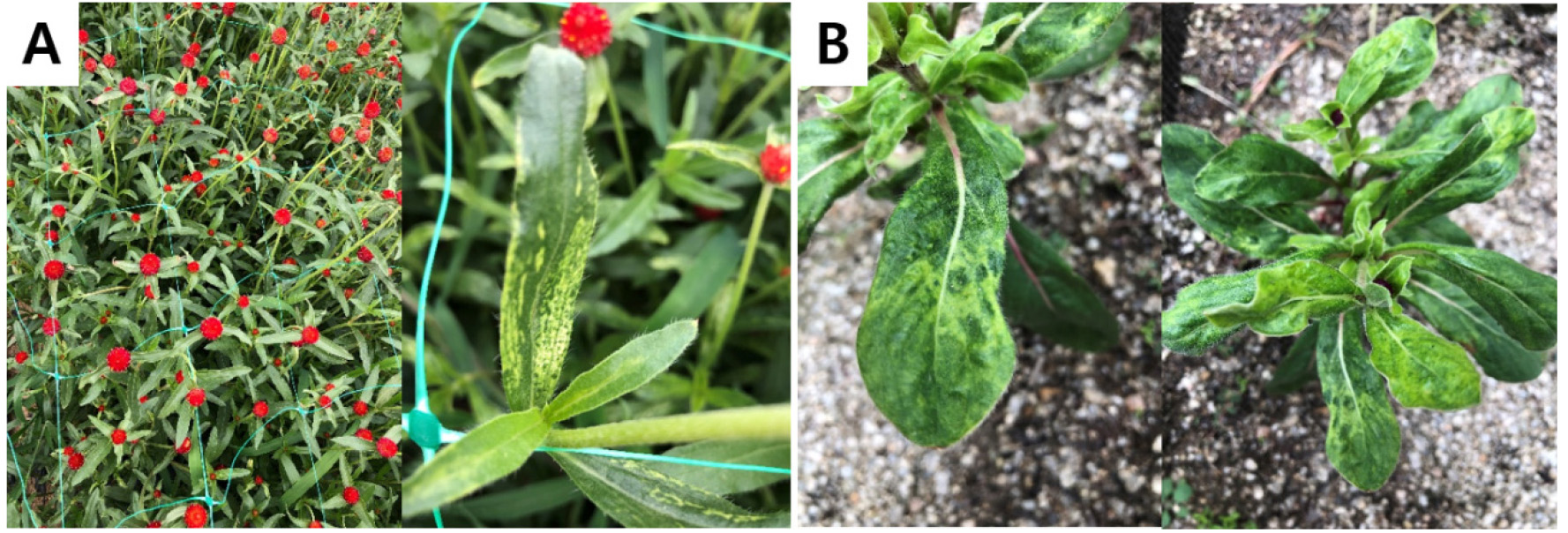
Fig. 1.
Mosaic symptoms observed on Gomphrena globosa naturally infected with cucumber mosaic virus (CMV). (A) Mosaic symptoms observed on the CMV-GomCC1 isolate collected from Chuncheon, Korea. (B) Mosaic symptoms observed on the CMV-GomCC2 isolate collected from a different location in Chuncheon, Korea.
The reverse transcription reaction was performed at 42°C for 1 h, followed by enzyme inactivation at 92°C for 5 min. PCR was repeated for 35 cycles in total, with each cycle consisting of 30 s at 95°C, 30 s at 52°C, and 1 min at 72°C. Virus testing was conducted using genus-specific primers (Table 1) targeting Ilarvirus, Potyvirus, Fabavirus, Orthotospovirus, Potexvirus, Tobamovirus, and Cucumovirus, including the genus of the virus previously reported in G. globosa belongs. Amplification products were verified by electrophoresis on a 1% agarose gel.
Table 1.
Primer list used for virus detection in Gomphrena globosa
| Virus | Primer name | Sequence (5’ → 3’)1) | Size (bp) | Reference |
| Ilarvirus | Ilar-5’ | GCNGGWTGYGGDAARWCNAC | 309 | Untiveros et al. (2010) |
| Ilar-3’ | AMDGGWAYYTGYTYNGTRTCACC | |||
| Fabavirus | Faba-genus-5’ | AAATATTAAAACAAACAGCTTTCGTT | 390 | Ferrer et al. (2007) |
| Faba-genus-3’ | TTCAAAGCTCGTGCCATITYATTKGC | |||
| Orthotospovirus | Tospovirus-5’ | TCIRDICKIYKRAAICTCMSRTC | 450 | Okuda and Hanada (2001) |
| Tospovirus-3’ | GGGGGAGAGCAATYGWGTCA | |||
| Potexvirus | Potexvirus-5’ | CAYCARCARGCMAARGAYGA | 580 | Van der Vlugt and Berendsen (2002) |
| Potexvirus-3’ | AGCATRGCISCRTCYTG | |||
| Potyvirus | Poty-CI-For | GGIVVIGTIGGIWSIGGIAARTCIAC | 700 | Ha et al. (2008) |
| Poty-CI-Rev | ACICCRTTYTCDATDATRTTIGTIGC | |||
| Tobamovirus | TobamodF | TKGAYGGNGTBCCNGGNTGYGG | 880 | Li et al. (2018) |
| TobamodR | ACNGAVTBNABCTGTAATTGCTAT | |||
| Cucumovirus | CPT-All-F | YASYTTTDRGGTTCAATTCC | 950 | Choi et al. (1999) |
| CPT-All-R | GACTGACCATTTTAGCCG |
Host Reaction Experiments
CMV-GomCC1 and CMV-GomCC2 were each purified by isolating single local lesions formed after sap inoculation of Chenopodium quinoa with symptomatic leaves from the original samples. The isolated single local lesions were subsequently inoculated onto Nicotiana tabacum ‘Xanthi nc’. Symptomatic upper leaves were subsequently used as the inoculum for host response experiments. The inoculum was prepared by grinding 1 g of symptomatic leaf with 1 mL of 0.01 M phosphate buffer (pH 7.2). The prepared inoculum was inoculated by evenly rubbing it onto the leaf surfaces of carborundum-treated plants (Table 2). Additionally, CMV-Fny, which was maintained for the long term in our laboratory, was utilized as a control isolate to compare the host responses of CMV-GomCC1 and CMV-GomCC2. It was mechanically inoculated onto the same host plants under the same conditions. The inoculated plants were grown in a growth chamber maintained at 25-27°C and 60% relative humidity, with a photoperiod set to 16 h of light and 8 h of darkness to observe symptom onset. For plants exhibiting no symptoms, total RNA was extracted from the upper leaves, and RT-PCR using CMV-specific primers was conducted to confirm infection.
Table 2.
Host response of cucumber mosaic virus (CMV)-GomCC1 and GomCC2
| Family | Host plants | Symptoms of the leaves1) | ||
| GomCC1 | GomCC2 | Fny2) | ||
| Solanaceae | Nicotiana benthamiana | -/M | -/M | -/M |
| N. tabacum ‘Xanthi nc’ | -/M | -/M | -/M | |
| N. glutinosa | -/M | -/M | -/M | |
| Capsicum annuum ‘Sinhong’ | -/M | -/M | -/M | |
| C. annuum ‘Baerotta’ | -/- | -/- | -/- | |
| C. annuum ‘Bukang’ | -/- | -/- | -/- | |
| Chenopodiaceae | Chenopodium quinoa | Nll/- | Nll/- | Nll/- |
| C. amaranticolor | Nll/- | Nll/- | Nll/- | |
| Cucurbitaceae | Cucumis sativus ‘Dancheong’ | -/mM | -/SL | -/M |
| Cucurbita moschata | -/SL | -/SL | nt | |
1)Inoculated/upper leaves; M, mosaic; Nll, necrotic local lesions; mM, mild mosaic; SL, symptomless; -, no infection; nt, not tested.
2)CMV-Fny was used as a control (Banik et al., 1983)
Sequence Analysis and Phylogenetic Analysis
RT-PCR was performed using the following primers to obtain the MP and CP gene sequences of CMV-GomCC1 and CMV-GomCC2:CMV-MP (Fw; 5’-ATG GCT TTC CAA GGT ACC AGT A-3’, Rv; 5’-CTA AAG ACC GTT AAC CAC CTG-3’), and CMV-CP (Fw; 5’-ATG GAC AAA TCT GAA TCA ACC AG-3’, Rv; 5’-TCA GAC TGG GAG CAC TCC A-3’). The amplified PCR products were inserted into the pGEM-T Easy TA cloning vector system (Promega, USA) and their nucleic acid sequences were determined by Macrogen (Seoul, Korea). The obtained MP and CP sequences were registered in GenBank; the MP and CP accession numbers of CMV-GomCC1 are LC915911.1 and LC915909.1, respectively, and those of CMV-GomCC2 are LC915912.1 and LC915910.1, respectively. The obtained nucleic acid sequences were compared with previously reported CMV isolates using NCBI BLASTn to verify homology. The subsequent sequence alignment and phylogenetic analysis were conducted using MEGA10.
A total of 19 CMV isolate sequences, including CMV-GomCC1 and CMV-GomCC2, were employed for the phylogenetic analysis, with peanut stunt virus (PSV-ER) designated as the outgroup. The nucleic acid sequences of the isolates used for comparison were obtained from the GenBank database. All sequences were aligned using ClustalW, and a phylogenetic tree was constructed using the maximum likelihood method and 1,000 bootstrap repetitions (Fig. 2). The obtained CP amino acid sequences were aligned using ClustalW along with the sequences of a CMV isolate derived from Iranian G. globosa (CMV-Go5, KP455738.1) registered in GenBank and representative isolates (CMV-Fny and CMV-Y). The aligned amino acid sequences were compared and visualized using BioEdit version 7.2.5 (Fig. 3).
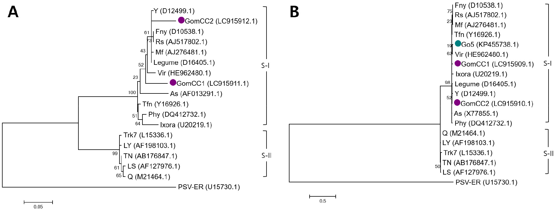
Fig. 2.
Phylogenetic analysis based on the amino acid sequences of the movement (A) and coat proteins (B) of cucumber mosaic virus (CMV)-GomCC1 and GomCC2, along with other CMV isolates including those derived from Gomphrena globosa. Phylogenetic analysis was performed using the maximum likelihood method with 1,000 bootstrap replications implemented in MEGA 10 following multiple alignment by ClustalW. CMV isolates were classified into three subgroups, and peanut stunt virus (PSV-ER) was used as the outgroup. The sequence of other CMV isolates were obtained from the GenBank database. The purple circle indicates the newly isolated CMV-GomCC1 and GomCC2 identified in this study.

Fig. 3.
Sequence alignment of the coat protein (CP) amino acid sequences of CMV isolates from globe amaranth. The CP amino acid sequences of the Korean Gomphrena globosa isolates (CMV-GomCC1 and CMV-GomCC2) were compared with the sequence of the Iranian G. globosa-derived isolate deposited in GenBank (CMV-Go5, KP455738.1) and representative CMV isolates (CMV-Fny and CMV-Y). The N-terminal arginine-rich motif is indicated above the alignment. Dots represent amino acids identical to those of CMV-GomCC1.
Results and Discussion
Gomphrena globosa plants showing mosaic symptoms were collected from two different locations in Chuncheon, Gangwon Province, South Korea and tested for viral infection. Total RNA was extracted from the leaves exhibiting symptoms, and RT-PCR testing was conducted for seven viral genera (Ilarvirus, Potyvirus, Fabavirus, Orthotospovirus, Potexvirus, Tobamovirus, and Cucumovirus) to which viruses reported in G. globosa belong (Table 1). Consequently, amplification products of the expected size (approximately 950 bp) were detected only with the primers specific to the genus Cucumovirus (data not shown). Sequence analysis of the amplified products confirmed that both samples were infected by the CMV, and the respective isolates were named CMV-GomCC1 and CMV-GomCC2. CMV-GomCC1 exhibited 99.85% nucleic acid sequence homology with a domestic Browallia speciosa isolate (Accession No. LC912011.1), whereas CMV-GomCC2 showed 99.24% nucleic acid sequence homology with a domestic Piper sarmentosum isolate (PQ790231.1).
Sap inoculation was performed on eight host plant species, including Nicotiana benthamiana, to assess the host responses of CMV-GomCC1 and CMV-GomCC2 (Table 2). Both isolates induced mosaic symptoms on the upper leaves of N. benthamiana, N. tabacum ‘Xanthi nc’, and N. glutinosa, and mosaic symptoms were also observed on Capsicum annuum ‘Sinhong’. In contrast, in C. annuum ‘Baerotta’ and ‘Bukang’, no symptoms were noted in either isolate, and CMV was not detected via RT-PCR. In Chenopodium quinoa and C. amaranticolor, necrotic local lesions formed on the inoculated leaves, whereas in Cucurbita moschata, although no visible symptoms appeared in either isolate, CMV infection was confirmed via RT-PCR, indicating symptomless infection. CMV-Fny, a representative CMV isolate, exhibited similar symptom patterns in most host plants. In particular, in Cucumis sativus ‘Dancheong’, CMV-Fny showed typical mosaic symptoms on the upper leaves, whereas CMV-GomCC1 exhibited a mild mosaic pattern. In contrast, CMV-GomCC2 did not show any symptoms, and RT-PCR analysis confirmed a symptomless infection. Thus, although the two CMV isolates derived from G. globosa generally exhibited host responses similar to those of CMV-Fny, differences exist in symptom manifestation in some hosts. Symptom manifestation after CMV infection can be influenced by different virus-host interactions, such as viral load, the host’s defense response, and the efficiency of intercellular or long-distance viral movement. These characteristics differ depending on the combination of viral isolate and host (Choi et al., 2005; Mochizuki and Ohki, 2012). Thus, the mild mosaic symptoms induced by CMV-GomCC1 in cucumber plants and the symptomless infection caused by CMV-GomCC2 in cucumber plants may be associated with the differences between the two isolates in viral load, host defense responses, or viral movement efficiency. However, given that this study only confirmed the infection status via RT-PCR, further analysis of viral load, movement characteristics, and host defense responses is required to identify the specific causes of these differences in symptoms. Meanwhile, RT-PCR testing of the original samples of seven viral genera detected no viruses other than Cucumovirus.
Phylogenetic analysis was conducted using the MP and CP amino acid sequences to confirm the phylogenetic positions of CMV-GomCC1 and CMV-GomCC2 (Fig. 2). Consequently, both isolates were confirmed to belong to CMV subgroup I and formed a distinct clade separate from subgroup II. In the MP phylogenetic tree, CMV- GomCC1 clustered closest to the LY2 isolate, whereas CMV-GomCC2 was positioned in the same clade as the Legume isolate (Fig. 2A). In contrast, in the CP phylogenetic tree, CMV-GomCC1 clustered close to the O isolate, whereas CMV-GomCC2 belonged to the same clade as the Y and GTN isolates (Fig. 2B). CMV subgroups are the primary taxonomic criteria for indicating phylogenetic relationships among isolates, and various biological characteristics may exist depending on the subgroup and isolate (Mochizuki and Ohki, 2012; Roossinck et al., 1999). In the present study, both CMV-GomCC1 and CMV-GomCC2 belonged to subgroup I and showed similar responses in most hosts. However, they exhibited different symptom expression patterns in cucumber. Thus, even CMV isolates belonging to the same subgroup may exhibit distinct biological characteristics in specific hosts, and phylogenetic subgroups alone may not adequately predict host responses or disease symptom expression.
Meanwhile, although no studies on the biological characteristics of CMV isolates derived from G. globosa have been reported, the CP sequence (KP455738.1) of CMV-Go5, derived from G. globosa in Iran, is registered in GenBank. Accordingly, the CP amino acid sequences of CMV-GomCC1 and CMV-GomCC2, isolated in Korea, were compared with those of CMV-Go5 and the representative isolates CMV-Fny and CMV-Y (Fig. 3). The sequence alignment results revealed that CMV-GomCC1 and CMV-GomCC2 showed high overall amino acid sequence conservation and shared mostly identical amino acid sequences with the isolate derived from the Iranian G. globosa. In contrast, the CP protein was highly conserved overall, with only a limited number of amino acid substitutions at specific positions compared to CMV-Fny and CMV-Y. In particular, the arginine-rich motif located at the N-terminus of CMV CP contributes to viral RNA binding and virus particle formation (Kaplan et al., 1998). Moreover, no amino acid substitutions were observed in this region among the CMV isolates derived from G. globosa compared in this study. Thus, despite some CP sequence variations among the G. globosa-derived isolates, the key functional regions associated with RNA binding and virus particle formation remain conserved.
This study examined the biological and molecular characteristics of the CMV isolates derived from Korean G. globosa. The two isolates were compared with the representative isolate, and differences in symptom expression were observed in some hosts. Additionally, the CP amino acid sequences among CMV isolates derived from G. globosa both domestically and internationally were highly conserved. These findings provide insights into the characteristics of CMV infecting G. globosa and serve as the foundational data for future studies on the pathogenicity and genetic diversity of CMV isolates derived from G. globosa.