


Introduction
Methodology
Plant Materials and Extraction Methods
Cell Culture
The 3-(4,5-dimethylthiazol-2-yl)-2,5-diphenyltetrazolium bromide (MTT) Assay
Microscopic Observations
Quantitative Real-time PCR (qPCR)
Statistical Analysis
Results and Discussion
Anti-proliferative Effects of the Sorghum Extracts on Human Cancer Cells
Observation of Morphological Disruption in Cancer Cells after Treatment with Sorghum Extracts
Effects of Sorghum Extract Treatment on the Expression of Apoptosis-related Genes
Summary
Introduction
Rapid industrialization, westernized lifestyle, and environmental pollution in modern society disrupt the body’s metabolic balance, thereby steadily increasing the incidence of cancer (Bray et al., 2024). Presently, in clinical practice, chemotherapy and radiation therapy are routinely employed together with surgery as the standard treatments for cancer. However, these physical and chemical therapies have a crucial limitation; they induce indiscriminate cytotoxicity in cancer and healthy cells, resulting in serious side effects such as hair loss, impaired immune function, cardiovascular disease, and multidrug resistance development (Bukowski et al., 2020). Consequently, global research efforts have recently been focused on identifying alternative anticancer compounds derived from natural plants that can maximize treatment efficacy by particularly targeting cancer cells while minimizing side effects on the human body (Garcia-Oliveira et al., 2021).
The most crucial strategy for developing successful natural anticancer agents is to inhibit the abnormal, uncontrolled proliferation of cancer cells and induce active cell death or apoptosis (Carneiro and El-Deiry, 2020). Apoptosis, an essential physiological mechanism for maintaining tissue homeostasis, is mainly regulated via two pathways: the extrinsic pathway, which involves cell surface receptors, and the intrinsic pathway, which is mitochondria-centered (Bertheloot et al., 2021). Of these, the balance in the expression of the BCL2 family of genes, which regulate the permeability of the mitochondrial outer membrane, determines the intrinsic pathway (Bock and Tait, 2020). When chemical or physical stress signals occur within cancer cells, BCL2 transcription, which inhibits apoptosis, reduces. Contrastingly, BAX expression, which promotes apoptosis, increases (Strasser and Vaux, 2020). Consequently, when cytochrome c is released from mitochondria, whose permeability has altered, into the cytoplasm, the early apoptosis signal transducer CASP9 is activated (Peng et al., 2022). Through a further reaction cascade, the final effector, CASP3, is activated. These activated enzymes block DNA repair mechanisms, accompanied by distinct morphological alterations such as cell shrinkage, chromatin condensation, and apoptotic body formation, which lead cancer cells to apoptosis (Asadi et al., 2022). Thus, the discovery of functional materials capable of precisely regulating this mitochondria-mediated apoptosis mechanism is crucial in tumor suppression research.
Meanwhile, sorghum is one of the world’s five major grains, which is widely cultivated mainly in Asia and Africa. It contains large amounts of secondary metabolites, such as polyphenols and flavonoids, with potent antioxidant activity (Khalid et al., 2022). The nutrients in sorghum help regulate the body’s glycemic response, which can decrease the risk of metabolic diseases such as diabetes, and are especially beneficial for people suffering from conditions such as celiac disease, diabetes, and obesity (Khoddami et al., 2023). Recently, sorghum has gained significant traction as its anticancer properties, which inhibit the cell cycle and induce apoptosis in cancer cells across different malignant tumor models, have been reported in succession (Chen et al., 2021; Santana et al., 2025). However, despite this high utility, studies that directly compare the anticancer efficacy among different collection resources with genetic diversity and those that specifically investigate the cell death mechanisms at the molecular level are scarce.
With three sorghum seeds with various genetic resources, IT225078, IT300108, and IT320916, we aimed to compare and examine the anti-proliferative effects on six human-derived cancer cell lines: AGS, A549, HCT116, HeLa, Huh7, and HepG2 and their cytomorphological changes. Additionally, we analyzed the changes in the mRNA expression of the BAX, BCL2, CASP3, and CASP9 after treatment with each extract to assess the effects of sorghum extracts on the expression of genes linked to mitochondria-dependent apoptosis.
Methodology
Plant Materials and Extraction Methods
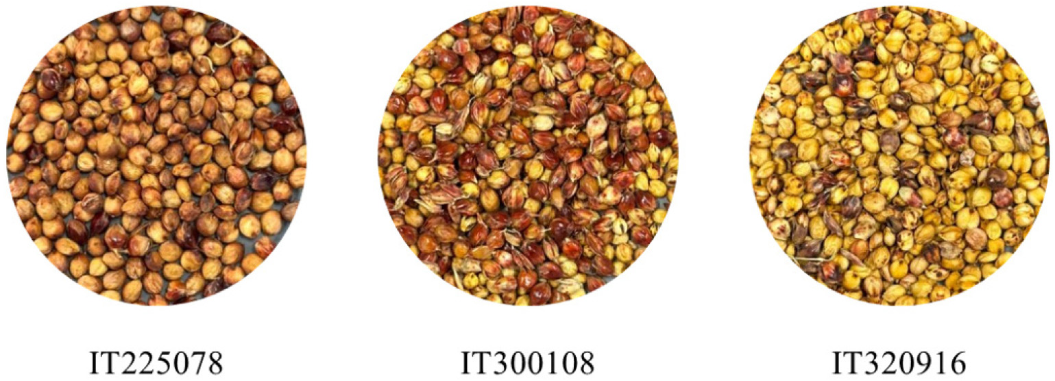
The three sorghum genotypes used in this experiment were all collected from the Gangwon Province region of South Korea. The specific collection sites for each resource were as follows: IT225078 from Sanae-myeon, Hwacheon-gun, Gangwon Province; IT300108 from Girin-myeon, Inje-gun, Gangwon Province; and IT320916 from Hyonnae-myeon, Goseong-gun, Gangwon Province. The seeds were obtained from the National Agrobiodiversity Center in 2023 and were further cultivated and harvested at the Kangwon National University Farm for use as experimental materials (Fig. 1). The seeds, dried following harvest, were ground into powder using a hand mixer (DH5001B, Korea). The prepared powder samples were mixed with 70% ethanol at a ratio of 1:10 (w/v) and subsequently soaked at room temperature for 72 h to perform the extraction. The extract was then filtered through filter paper (HYUNDAI MICRO NO. 20, HYUNDAI MICRO, Korea) to remove impurities, and the resulting filtrate was concentrated under reduced pressure at 40°C using a rotary evaporator (EYELA N-1110, Tokyo, Japan). The final concentrated extract was diluted to a 100,000 µg/mL concentration for further use in this study.
Cell Culture
The six human cancer cell lines used in this experiment (AGS, A549, HCT116, HeLa, HepG2, and Huh7) were obtained from the Korean Cell Line Bank (KCLB, Seoul, Korea). RPMI-1640 medium (Hyclone) was utilized as the basal medium for cell culture, to which 10% fetal bovine serum (FBS; Welgene, Gyeongsan, Korea) and 1% penicillin/streptomycin (Thermo Fisher Scientific, Waltham, MA, USA) were supplemented. Cells were cultured in an incubator (Sanyo Co., Ltd., MCO-19AIC, Osaka, Japan) maintained at a constant condition of 37°C temperature and 5% CO2. Cells were subcultured every 2-3 days once they reached 80%-90% cell density to ensure optimal growth conditions and physiological stability.
The 3-(4,5-dimethylthiazol-2-yl)-2,5-diphenyltetrazolium bromide (MTT) Assay
The MTT assay was conducted according to the method previously described (Hwang et al., 2022) to assess the anti-proliferative effects of the sorghum extract on cancer cells. The six previously cultured human cancer cell lines were seeded at a density of 5 × 10⁴ cells/well in 100 µL per well into a 96-well plate (SPL Life Sciences Co., Ltd., Pocheon, Korea) and were allowed to stabilize for 24 h in an incubator at 37°C and 5% CO2. The control group consisted of cells cultured in medium without any extract. This group was used as the reference (100% cell viability) to calculate the relative cell viability of each treatment group. Next, the existing medium was removed, and sorghum extract was diluted in a culture medium (RPMI-1640) to final concentrations of 25, 50, and 100 µg/mL. Furthermore, 100 µL of each solution was added to each well, followed by an additional 24 h of culture. The supernatant from the completed culture was carefully removed, and 100 µL of a 500 µg/mL solution of MTT was added to each well. The samples were subsequently allowed to react for 4 h under the same culture conditions. After the reaction, the supernatant was completely removed, and 100 µL of dimethyl sulfoxide was added to each well to dissolve the formazan crystals formed by reduction, followed by incubation at room temperature for 20 min to promote dissolution. Finally, absorbance was measured at 519 nm wavelength using a UV-Vis spectrophotometer (Thermo Fisher Scientific Inc.), and cell viability was calculated according to the following equation:
Microscopic Observations
Microscopic observations were conducted to investigate the morphological alterations in human cancer cells after treatment with sorghum extract. The six previously cultured cancer cell lines were seeded into a 6-well plate at a density of 1 × 10⁶ cells/well and cultured in an incubator until the cell density reached at least 80%. Subsequently, the existing medium was removed, and the cells were washed with 1× phosphate-buffered saline (PBS). Subsequently, RPMI-1640 medium (Hyclone) containing 100 µg/mL of sorghum extract was added to each well. After additional incubation for 24 h, the cells were examined using a microscope to check for morphological alterations such as cell atrophy, detachment, and cell membrane damage. The images were acquired and the observed cells were analyzed using iSolutionLite (IMT i-solution Inc., Quebec, Canada).
Quantitative Real-time PCR (qPCR)
We confirmed the changes in the expression of apoptosis-related genes after treatment with sorghum extract by performing qPCR according to the method previously described (Seo et al., 2023). First, TRIzol reagent (Thermo Fisher Scientific Inc.) was used to extract the total RNA from each cell treated with sorghum extract, and the quantity and purity of the extracted RNA were measured using a microvolume spectrophotometer (Keen Innovative Solutions, Seoul, Korea). The quantified RNA was reverse-transcribed into cDNA using PrimeScriptTM RT Master Mix (Perfect Real Time; Takara Korea Biomedical Inc., Seoul, Korea) according to the manufacturer’s instructions. The qPCR reaction mixture was prepared to a total volume of 25 µL using TB Green® Premix Ex TaqTM (Tli RNaseH Plus; Takara Korea Biomedical Inc.), and gene amplification and real-time monitoring were conducted using the CronoSTARTM 96 Real-Time PCR System (Takara Korea Biomedical Inc.). The PCR amplification conditions consisted of an initial denaturation step at 95°C for 30 s, followed by 40 cycles of denaturation at 95°C for 5 s, and annealing and extension at 60°C for 30 s. After the amplification reaction was completed, a melting curve analysis (1 min at 95°C, 15 s at 60°C, and 5 s at 98°C) was conducted to check for the amplification of nonspecific products. Herein, the expression levels of BCL2, BAX, CASP3, and CASP9 were examined to identify the mechanisms of cancer cell death, in which GAPDH was used as an internal control for expression normalization. Relative mRNA expression levels were calculated using the 2-ΔΔCt method. Before analysis, the amplification efficiency and melting curves of the primers were verified to confirm single-product amplification. Additionally, the stability of GAPDH expression was confirmed to improve the reliability and reproducibility of the results. Table 1 shows the nucleotide sequences of all primers used in the experiment.
Table 1.
Primer sequences used for quantitative polymerase chain reaction analysis
Statistical Analysis
All experiments in this study were independently conducted at least three times, and the obtained results are expressed as the mean ± standard deviation. Statistical analysis was conducted using IBM SPSS Statistics (version 29). The qPCR differences between each extract-treated group and the control were analyzed using independent Student’s t-tests to align with the independent comparison objectives of the experimental design. Conversely, data needing comparisons among multiple groups, such as cell viability, were examined using one-way analysis of variance followed by Duncan’s multiple range test. Statistical significance was set at p < 0.05.
Results and Discussion
Anti-proliferative Effects of the Sorghum Extracts on Human Cancer Cells
The MTT assay is a representative technique for assessing cytotoxicity based on the principle that mitochondrial dehydrogenase in living cells decreases the yellow MTT reagent to form purple formazan crystals (Benov, 2021). Herein, we evaluated the anti-proliferative effects of ethanol extracts from three sorghum genetic resources (IT225078, IT300108, and IT320916) collected in Gangwon Province against six human cancer cell lines (AGS, A549, HCT116, HeLa, HepG2, and Huh7).
The treated sorghum extracts generally showed anti-proliferative effects against cancer cells in a concentration- dependent manner (Fig. 2). Particularly, the IT320916 extract recorded the lowest cell viability in most of the six cancer cell lines, including the AGS and A549 cell lines, at the highest treatment concentration of 100 µg/mL. This further revealed the strongest anticancer activity compared to the other resources, IT225078 and IT300108. This suggests that the IT320916 resource has the potential as an excellent anticancer functional material. When treated with extracts containing IT320916, the cell viability of Huh7 cells exhibited a concentration-dependent reduction. However, even in the high-concentration treatment group (100 µg/mL), the cell viability remained over 70%, indicating a relatively minor cytotoxic effect compared to the other cancer cell lines (Fig. 2E).
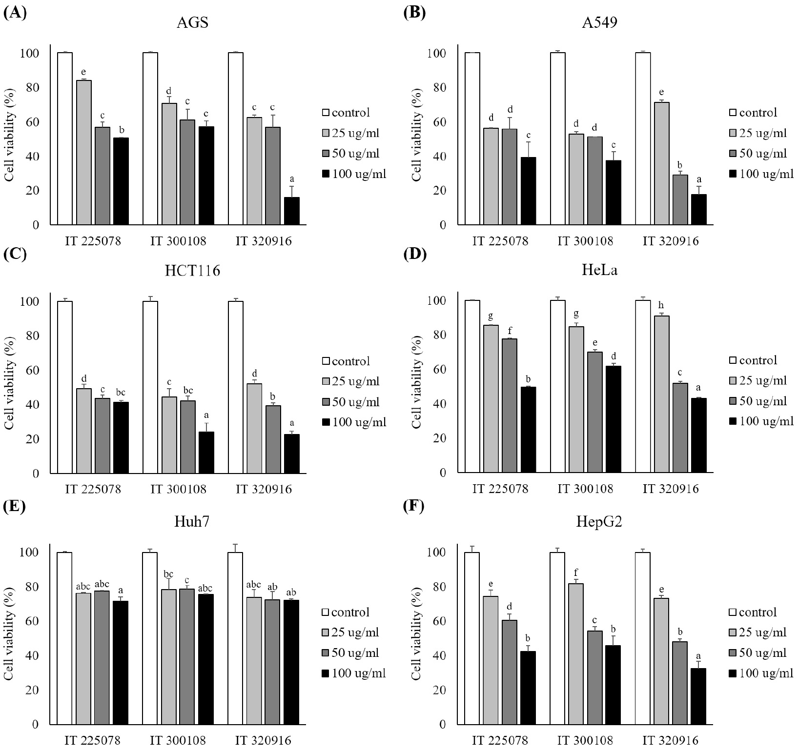
Fig. 2.
Inhibitory effects of the sorghum extracts on the proliferation of different human cancer cells. Cell viability of (A) AGS, (B) A549, (C) HCT116, (D) HeLa, (E) Huh7, and (F) HepG2 cells was assessed using the MTT assay following treatment with 25, 50, 100 µg/mL of the extracts for 24 h. Untreated vehicle control was set to 100% cell viability. Error bars represent the standard deviation (SD) of three independent biological replicates (n=3). Different letters indicate the statistically significant differences among all groups including the control (one-way analysis of variance followed by Duncan’s multiple range test, p < 0.05).
These findings are consistent with those of previous studies, which revealed that phenolic compounds contained in sorghum effectively inhibited cancer cell proliferation. Xu et al. (2021) reported that polyphenolic compounds in sorghum can act as tumor suppressors in different malignant tumor models (Xu et al., 2021). Seo et al. (2025) revealed that different sorghum seed extracts that are rich in phenolic compounds showed potent anticancer activity by regulating the expression of apoptosis-related genes (Seo et al., 2025). This is consistent with the findings of this study, which showed excellent anticancer activity using sorghum extract. Additionally, the efficacy of the sorghum extract used in this study is especially notable when compared to the extracts from other medicinal plants. Teruna et al. (2026) reported that when Lithocarpus bancanus extract was applied to the A549 cells, the IC50 values ranged from 304.7 to 455.1 µg/mL depending on the extraction solvent (Teruna et al., 2026). Notably, the IT320916 extract used in this study showed cytotoxic activity against most cancer cells even at a 100 µg/mL concentration, which is 3 to 4 times lower than those concentrations in the previous study. Additionally, its efficacy against the colorectal cancer cell line HCT116 is remarkable. Rahamooz-Haghighi et al. (2021) reported an IC50 value of 897.14 µg/mL for HCT116 cells after a 24-h treatment with an ethanol extract of Plantago major roots. The extract used in this study showed outstanding anti-proliferative effects at 100 µg/mL concentration, which is only one-eighth of that level (Rahamooz-Haghighi et al., 2021). This indicates that the sorghum extract is a highly effective functional material that shows outstanding cytotoxic effects even at relatively low concentrations compared to the other known anticancer agents. Collins et al. (2026) reported that a phenolic extract containing sorghum pigments effectively induced apoptosis by controlling the expression of key genes linked to cancer development pathways (Collins et al., 2026).
Thus, the potent cytotoxicity observed in this study is believed to result from the high concentrations of antioxidants and bioactive compounds present in the sorghum extract. This disrupts the signaling pathways of cancer cells, thereby inhibiting their abnormal proliferation. In conclusion, sorghum extracts from genetic resources, especially from IT320916, have high potential as excellent preventive and therapeutic agents against different cancer types, including AGS and A549. Thus, in-depth molecular biological research should continue to elucidate the specific mechanisms underlying apoptosis induction.
Observation of Morphological Disruption in Cancer Cells after Treatment with Sorghum Extracts
Six cancer cell lines were treated with the extracts at a concentration of 100 µg/mL for 24 h, followed by microscopic observation to investigate the effects of sorghum extract on cancer cell viability and on actual morphological changes in the cells (Fig. 3). Before microscopy, the samples were washed with 1× PBS to remove suspended culture medium components and debris from dead cells.
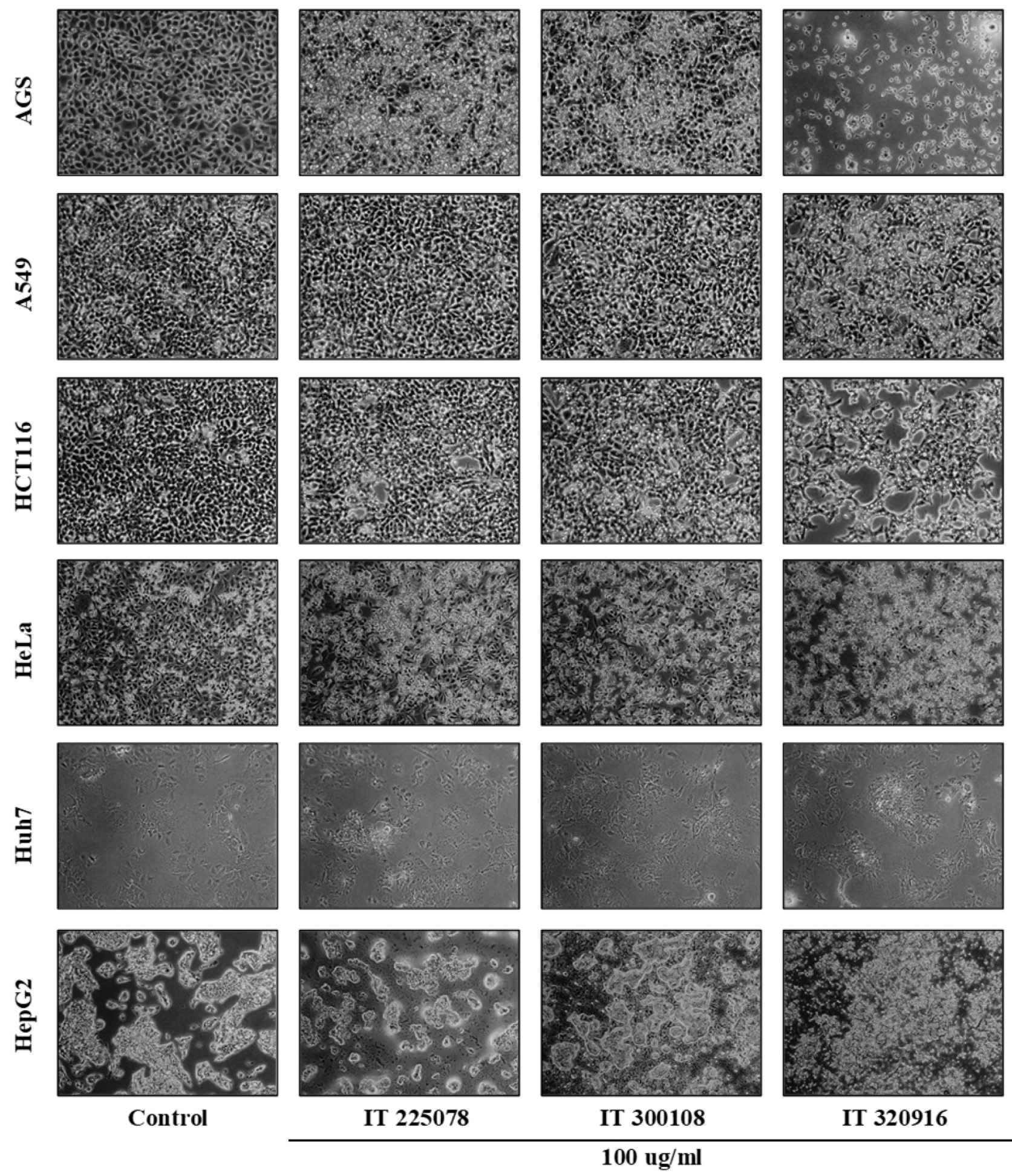
Fig. 3.
Microscopic observation of the cellular morphological changes induced by the sorghum extracts in human cancer cell lines. Cells were treated with 100 µg/mL of the extracts for 24 h, and morphological changes, such as cell shrinkage and detachment, were observed under a phase-contrast microscope.
The six cancer cell lines, including the control AGS and A549, maintained irregular polygonal or epithelial cell shapes, adhering firmly to the bottom of the culture dishes and proliferating at high densities. These findings are consistent with the morphological characteristics of cancer cells identified in a previous study (Micucci et al., 2025). The experimental groups treated with sorghum extract showed a substantial loss of morphological integrity compared to the control group. In the groups treated with IT225078 and IT300108 extracts, some cells lost their original shape and became rounded and atrophied. However, these morphological changes and cell death signs were the most pronounced in the group treated with the IT320916 extract. In the case of the gastric cancer cell line AGS, the cell density reduced most rapidly upon treatment with the IT320916 extract. Generally, when cell death is induced, the cell cytoskeleton disintegrates, causing the cells to round up and shrink in volume, eventually detaching from the culture vessel surface (Wani et al., 2023). Additionally, this may be attributed to the loss of cell adhesion and subsequent detachment caused by the extract’s potent toxicity (Mokoena et al., 2022), resulting in their removal during the previous PBS wash step. Furthermore, the majority of cells remaining in the culture vessels were severely atrophied and deformed into round shapes, having lost their normal cell membrane morphology. Thus, the pronounced cell shrinkage observed in the IT320916-treated group, compared to the partial shrinkage in the IT225078 and IT300108 groups, provides morphological evidence that the IT320916 extract has the most potent cell death-inducing activity. The liver cancer cell line Huh7 exhibited varying outcomes. Despite treatment with sorghum extracts, including IT320916, which showed the strongest activity, at a concentration of 100 µg/mL, the majority of cells adhered to the culture dishes, similar to the normal control group. Additionally, almost no cell atrophy or morphological damage was noted. This is consistent with the findings of the previous MTT assay, in which Huh7 cells showed a relatively high cell viability compared to other cancer cells. This is likely because Huh7 cells has a mutant p53 protein (Bressac et al., 1990).
In summary, sorghum extracts, especially IT320916, caused cell membrane damage and morphological disruption in the AGS, A549, HCT116, HeLa, and HepG2 cells. However, Huh7 cells were relatively less susceptible and exhibited only a negligible effect.
Effects of Sorghum Extract Treatment on the Expression of Apoptosis-related Genes
Changes in the mRNA expression of the key apoptosis regulators were analyzed via qPCR to elucidate the mechanism of cancer cell death induced by sorghum extract treatment (Fig. 4). Generally, mitochondria-dependent cell death begins when p53 and other factors increase the expression of the intracellular transcription activator BAX and suppress the expression of the anti-apoptotic factor BCL2. Consequently, mitochondrial membrane permeability changes, resulting in cytochrome c releasing into the cytoplasm. This activates CASP9, which ultimately triggers a cascade of CASP3 activations, leading to cell death (Sarman and Asci, 2026).
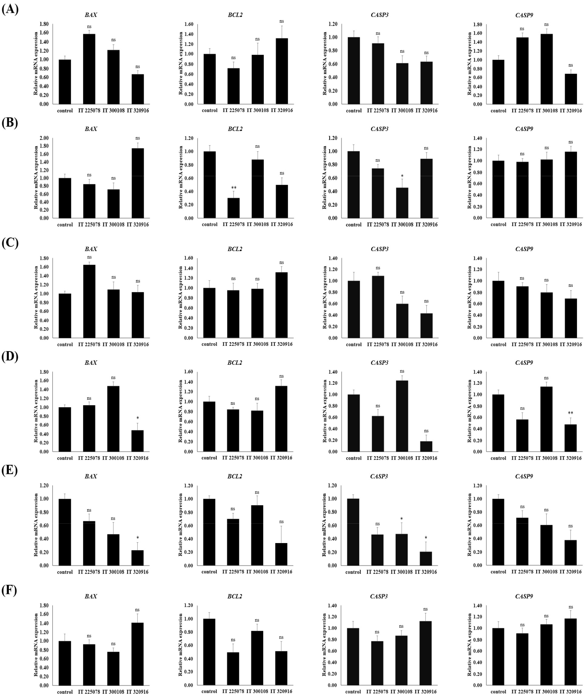
Fig. 4.
Modulatory effects of the sorghum extracts on the transcriptional levels of apoptosis-related genes. The relative mRNA expression of BAX, BCL2, CASP3, and CASP9 was determined by quantitative polymerase chain reaction in (A) AGS, (B) A549, (C) HCT116, (D) HeLa, (E) Huh7, and (F) HepG2 cells treated with 100 µg/mL of the extracts for 24 h. GAPDH was used as an internal control for normalization. Error bars represent the standard deviation (SD) of three independent biological replicates (n=3). Statistical significance comparing each extract treatment exclusively to the untreated control was determined using Student’s t-test (*p < 0.05; p < 0.01; ns, not significant).
The analysis showed that the expression patterns of apoptosis-related genes after treatment with sorghum extracts differed depending on the cell line and extract type. Since the qPCR analysis in this study revealed no statistically significant differences in the expression levels among the extract-treated groups, the effects of each extract on the expression of apoptosis-related genes were individually assessed compared to the untreated control group. Overall, in AGS and HCT116 cells, treatment with the IT225078 extract led to the upregulation of the BAX and Caspase family genes compared to the control group, whereas BCL2 tended to be downregulated. This suggested that these extracts have the potential to participate in apoptosis mechanisms in gastrointestinal cancer cells. However, some results were inconsistent with this typical apoptotic activation pattern, depending on the cell line and extract type. For instance, alterations in gene expression were not consistent across all conditions. CASP3 expression was significantly reduced after IT300108 treatment in the A549 cells, and the expression of BAX and CASP9 reduced in the HeLa cells after IT320916 treatment. Meanwhile, in the Huh7 cells, high-concentration treatment unexpectedly reduced the expression of apoptosis-related genes or maintained levels comparable to the control. Particularly, this low responsiveness of Huh7 cells is consistent with the high cell viability identified in the previous MTT assay and morphological observations and supports the relative resistance of this cell line to the sorghum extracts.
This resistance of Huh7 cells is presumed to be due to the inherent genetic characteristics of the cell line, especially the differences in the status and mechanisms of the tumor suppressor gene p53. Previously, based on an analysis of liver cancer cell lines, Bressac et al. (1990) reported that while HepG2 was the only line to have a normal wild-type p53 gene, Huh7 had a missense mutation (Y220C) characterized by abnormal p53 protein accumulation (Bressac et al., 1990). Bao et al. (2025) showed that the TP53 pathway plays a primary role when natural polyphenols regulate the BAX/BCL2 ratio in liver cancer cells and affect CASP3/9 expression to induce apoptosis (Bao et al., 2025). While normal p53 plays a critical role in recognizing cellular damage and triggering the cell death process, this mechanism may not function properly when p53 is functionally lost, as in the Huh7 cells (Tseng et al., 2022). Thus, the substantial cell death rate observed in HepG2 cells and the minimal changes in gene expression in the Huh7 cells in this study suggest that the anticancer mechanism of the sorghum extracts is linked to the functional activation of p53.
Meanwhile, the MTT assay and morphological observations in this study revealed that the IT320916 extract showed the strongest cytotoxicity in most cancer cell lines. However, qPCR analysis did not reveal any significant statistical differences in the expression levels of apoptosis-related genes among the three extracts. This implies that the mechanism by which the sorghum extracts induce cancer cell death may not be limited to a single pathway. In other words, while all three resources affected the expression of mitochondria-dependent apoptosis-related genes to a similar extent, the IT320916 resource, which showed the most potent cytotoxicity, likely activated a combination of other cell death mechanisms in addition to apoptosis, such as necrosis caused by cell membrane disruption, autophagy, or the extrinsic pathway. Indeed, morphological observations revealing prominent rapid cell detachment and membrane damage in the IT320916 extract-treated group, support this hypothesis. Additionally, since the sorghum extracts are complex mixtures composed of different bioactive substances, such as polyphenols and tannins, rather than a single component, the active ingredients and the signaling pathways activated may vary depending on the genetic background and receptor expression characteristics of each cancer cell line (Liu, 2004).
Furthermore, since this study examined only the mRNA expression levels of the Caspase family, limitations exist in interpreting these results as direct evidence of protein cleavage or actual enzyme activation. Thus, the extracts from the sorghum genetic resources (IT225078, IT320916, and IT300108) acted specifically depending on the cancer cell type and exerted complex effects on the BAX/BCL2 ratio and the mRNA expression of the Caspase family genes. This suggests that the sorghum extracts exerts anticancer effects by participating in mitochondria- dependent cell death pathways. However, further investigation at the protein level, such as via western blot analysis, is necessary to confirm whether these mechanisms are actually activated.
Summary
This study assessed the anti-proliferative effects and molecular mechanisms of cell death in six human cancer cell lines using extracts from three sorghum genetic resources: IT225078, IT300108, and IT320916. The sorghum extracts decreased cancer cell viability in a concentration-dependent manner and induced morphological signs of typical cell death, such as cell shrinkage and detachment. Furthermore, gene expression analysis showed that the extract-treated groups demonstrated changes in the mRNA expression of BCL2, BAX, CASP3, and CASP9, suggesting a potential involvement in the expression of genes associated with mitochondria-dependent apoptosis. Meanwhile, relatively strong resistance was noted in the Huh7 cells harboring mutant p53. This suggested that the anticancer mechanism of the sorghum extracts is closely linked to the functional activation of p53. In conclusion, this study showed that sorghum extract can decrease the viability of different cancer cell lines and affect the mRNA expression of apoptosis-related genes, suggesting its potential as a natural product-based functional ingredient. If further supported by the isolation and identification of key bioactive components, along with additional validation of the anticancer mechanism at the protein level and under in vivo conditions, our results will serve as a crucial foundation for developing natural anticancer agents and high-value functional foods.