


Introduction
Materials and Methods
Plant Materials and Genomic DNA Extraction
Amplification, Cloning, and Sequence Analysis of SlGAD3
Design of Allele-Specific Primers and TaqMan Probe
Design and Construction of the Sequence-Traceable Positive-Control Plasmid
Sequence Verification of the Positive-Control Plasmid
Conventional PCR and Serial-Dilution Analysis
Specificity Analysis
TaqMan Real-Time PCR Analysis
Sequence Discrimination of the Positive-Control Amplicon
Data Analysis
Results
Molecular Confirmation of the Target SlGAD3 Sequence Variant in GABA-Enriched Tomato
Construction and Sequence Verification of the Sequence-Traceable Positive-Control Plasmid
Conventional PCR Reactivity of the Positive-Control Plasmid
TaqMan Real-Time PCR Amplification of the Positive-Control Plasmid
Copy-Number-Based Standard-Curve Characteristics of the TaqMan Real-Time PCR
Specificity of the SlGAD3_var1 Conventional PCR Assay
Discussion
Conclusion
Introduction
γ-Aminobutyric acid (GABA) is a four-carbon non-protein amino acid produced through the irreversible decarboxylation of L-glutamate by glutamate decarboxylase (GAD). In plants, GABA is involved in diverse physiological and metabolic processes, including carbon and nitrogen metabolism, intracellular pH regulation, stress responses, defense, and cellular signaling. GABA metabolism is also connected to the tricarboxylic acid cycle through the GABA shunt, which principally comprises reactions catalyzed by GAD, GABA transaminase, and succinic semialdehyde dehydrogenase. In addition to its metabolic role, GABA has been increasingly recognized as an important signaling molecule involved in the regulation of plant growth and responses to environmental stimuli (Bouché and Fromm, 2004; Bown and Shelp, 1997; Fait et al., 2008; Michaeli and Fromm, 2015; Shelp et al., 1999). Tomato (Solanum lycopersicum L.) accumulates relatively high concentrations of GABA during fruit development, particularly before the breaker stage. The tomato genome contains five GAD genes, designated SlGAD1-SlGAD5, among which SlGAD2 and SlGAD3 are prominently expressed during fruit development and play major roles in regulating GABA accumulation. Most plant GAD proteins contain a C-terminal regulatory region that includes a calmodulin-binding domain and functions as an autoinhibitory domain. Removal or disruption of this region can increase constitutive GAD activity and consequently enhance GABA accumulation. Expression of a C-terminally truncated SlGAD3 was shown to markedly increase GABA levels in tomato fruit, confirming the regulatory importance of the C-terminal autoinhibitory region (Akihiro et al., 2008; Takayama and Ezura, 2015; Takayama et al., 2017).
Targeted mutations were introduced into the C-terminal regions of SlGAD2 and SlGAD3 using the CRISPR/Cas9 system. Mutations that generated premature stop codons resulted in truncation of the autoinhibitory region and substantially increased GABA accumulation in tomato fruit. This approach demonstrated that targeted modification of a small endogenous regulatory sequence could generate a nutritionally enhanced crop phenotype without requiring the stable expression of a foreign GAD transgene. The resulting genome-edited GABA-enriched tomato therefore represents an important example of crop improvement through targeted mutagenesis of an endogenous metabolic gene (Nonaka et al., 2017).
The molecular characteristics of genome-edited crops present specific challenges for their molecular detection. Conventional genetically modified crops commonly contain introduced promoters, terminators, selectable marker genes, or plant-transgene junction sequences that can be detected using element-, construct-, or event-specific PCR assays. By contrast, a genome-edited crop may differ from its conventional counterpart by only a single-nucleotide substitution or a short insertion or deletion in an endogenous gene. General screening assays targeting frequently used genetic elements, such as the cauliflower mosaic virus 35S promoter or the Agrobacterium tumefaciens nopaline synthase terminator, cannot directly detect such edited alleles. Detection of the edited SlGAD3 allele in GABA-enriched tomato therefore requires sequence-specific analysis of the modified SlGAD3 region and sufficient discrimination between the edited sequence and the corresponding non-edited allele (Grohmann et al., 2019; Holst-Jensen et al., 2003). PCR-based methods targeting genome-editing sites have been developed for several genome-edited crops. A real-time PCR method was developed for a commercialized genome-edited canola line, and an editing-site-specific PCR method was established for the detection and quantification of CAO1-edited rice (Chhalliyil et al., 2020; Zhang et al., 2021). These studies demonstrated that genome-edited plants carrying short sequence variants can potentially be detected when the exact nucleotide sequence of the edited site is known. Nevertheless, distinguishing an edited allele from closely related wild-type alleles or naturally occurring variants remains analytically challenging, particularly when the sequences differ by only one or a few nucleotides (Chhalliyil et al., 2020; Zhang et al., 2021). Importantly, detection of the edited SlGAD3 sequence does not by itself demonstrate that the detected allele originated from CRISPR/Cas9-mediated genome editing. An identical endogenous sequence variant could potentially occur naturally or arise through another mutagenic process. Accordingly, the present assay is intended to detect the defined edited SlGAD3 allele rather than to identify the genome-editing process or a specific genome-edited event.
Allele-specific PCR is widely used to discriminate single-nucleotide polymorphisms and short insertion-deletion variants. In this approach, a nucleotide specific to the target allele is generally positioned at or near the 3’ terminus of an allele-specific primer, thereby reducing extension from mismatched non-target templates. The amplification refractory mutation system demonstrated that the discriminatory capacity of allele-specific PCR could be improved by controlling mismatches at the primer 3’ end. Additional deliberate mismatches introduced near the 3’ terminus have subsequently been used to enhance allelic discrimination, although mismatch number, position, and nucleotide combination must be optimized to avoid non-target or pseudo-positive amplification (Bui and Liu, 2009; Newton et al., 1989; Yaku et al., 2008). In the present detection system, SlGAD3_var1_F was used as the allele-specific forward primer for the edited SlGAD3 sequence. The designations “var1” and “var2” refer to alternative primer designs containing different deliberate mismatches and do not denote distinct biological SlGAD3 variants. Both primer designs targeted the same single-thymine insertion in the C-terminal autoinhibitory region of SlGAD3, identified relative to the reference sequence NM_001246898.2. This primer contained a deliberate mismatch intended to improve discrimination of the edited allele, and it was combined with the SlGAD3_WT_R reverse primer for conventional PCR and with the GABA-TaqMan probe for real-time PCR. Because the assay targets a short endogenous sequence variant in a tomato gene, the availability of a stable and well-characterized positive-control material is particularly important for method development, analytical validation, and routine application.
A sequence-defined amplification positive control can verify the performance of the polymerase, primers, probe, reaction mixture, thermal cycling, and signal-detection steps. However, a purified plasmid control does not assess DNA extraction efficiency, sample DNA integrity, or PCR inhibition associated with the tomato matrix and therefore should not be interpreted as an extraction control or internal amplification control (Caasi et al., 2013; Smith et al., 2006). Although authentic GABA-enriched tomato genomic DNA contains the edited SlGAD3 allele in its natural genomic context, its availability, quality, and long-term reproducibility may be limited. A plasmid containing a defined target sequence therefore provides a stable and renewable alternative for routine amplification control.
A major limitation of plasmid-based positive controls is the risk of carryover contamination. Because conventional control plasmids generally contain the same assay target as the authentic sample, plasmid-derived and genomic amplicons may be indistinguishable by agarose gel electrophoresis or routine TaqMan fluorescence analysis. To facilitate investigation of suspected positive-control carryover, traceable control constructs can incorporate artificial internal sequence features while retaining the primer- and probe-binding sites required for routine amplification. For example, the pBT63 positive-control plasmid incorporated a discriminatory restriction site that allowed control-derived amplicons to be distinguished from authentic products (Minegishi et al., 2014). In the present strategy, plasmid-specific spacer sequences were introduced as post-amplification identifiers. These sequences do not prevent contamination; rather, when carryover is suspected, Sanger sequencing of the amplicon can determine whether the product contains the plasmid-specific spacer sequences and therefore originated from the positive-control plasmid.
Therefore, the objective of the present study was to develop and analytically characterize a sequence-traceable positive-control plasmid for conventional PCR and TaqMan real-time PCR detection of the edited SlGAD3 allele in GABA-enriched tomato. The assay was designed to detect the defined SlGAD3 sequence variant rather than to establish the genome-editing process by which the variant arose. The plasmid retained the SlGAD3_var1_F-, GABA-TaqMan probe-, and SlGAD3_WT_R-binding sites while incorporating two plasmid-specific spacer sequences within the amplified region. The construct was evaluated by Sanger sequencing, conventional PCR, and TaqMan real-time PCR to characterize its amplification performance and determine whether plasmid-derived amplicons could be distinguished from the corresponding tomato genomic sequence.
Materials and Methods
Plant Materials and Genomic DNA Extraction
Sicilian Rouge High GABA tomato was commercially purchased in Japan from Sanatech Seed Co., Ltd. and received as freeze-dried fruit tissue. The presence of the target SlGAD3 sequence variant in this material was subsequently confirmed by PCR amplification, cloning, and Sanger sequencing as described below. Commercial Micro-Tom seeds were purchased from Xplant (Republic of Korea) and used as a sequence comparator for characterization of the SlGAD3 locus. Micro-Tom was not considered an isogenic parental line or the genetic background from which the GABA-enriched tomato was derived. The zygosity of the edited SlGAD3 allele in the commercial GABA-enriched tomato material analyzed in this study was not independently determined. In addition, 11 conventional commercial tomato samples were purchased through commercial retail channels in the Republic of Korea, including online retailers, Costco, and E-Mart, and were included in the specificity assessment. The commercial panel comprised Apple Jujube Tomato, Royal Gold Tomato, Orange Jujube Cherry Tomato, Black Jujube Cherry Tomato, Jujube Cherry Tomato, Yellow Gourd Cherry Tomato, Shine Cherry Tomato, The Hard Jujube Cherry Tomato, Ruby Bell Tomato, Cocktail Tomato, and Mini Chal Tomato. The samples were identified according to the cultivar or product names provided by the retailers at the time of purchase. Their GM or genome-editing status was not independently verified; therefore, they are referred to as conventional commercial tomato samples rather than non-GM or non-edited tomatoes. Lot or batch information was not recorded at the time of purchase. Plant materials used for DNA extraction consisted of freeze-dried fruit tissue from the GABA-enriched tomato, fruit flesh from Micro-Tom plants grown from commercially purchased seeds, and fruit tissue from the 11 conventional commercial tomato samples. Fresh fruit tissues were pulverized under liquid nitrogen before genomic DNA extraction. Genomic DNA was extracted using the APrepTM Seed & Bulb DNA Extraction Kit (APBIO, Republic of Korea) according to the manufacturer’s instructions. DNA concentration and purity were determined by UV-visible spectrophotometry, and DNA integrity was assessed by agarose gel electrophoresis. Extracted DNA was stored at -20°C until use.
Amplification, Cloning, and Sequence Analysis of SlGAD3
The full-length SlGAD3 region was amplified from genomic DNA obtained from GABA-enriched tomato and Micro-Tom using the SlGAD3_F and SlGAD3_R primer set. The expected size of the amplified fragment was 1,925 bp. High-fidelity PCR was performed using Q5® Hot Start High-Fidelity 2× Master Mix (New England Biolabs, USA). The amplification conditions consisted of initial denaturation at 98°C for 30 s, followed by 35 cycles of denaturation at 98°C for 10 s, annealing at 60°C for 30 s, and extension at 72°C for 2 min 30 s, with a final extension at 72°C for 2 min. The amplified products were separated by agarose gel electrophoresis and purified. Because amplification with Q5 high-fidelity polymerase generates blunt-ended products, the purified amplicons were subjected to A-tailing before ligation into the pMD20 T-vector. Recombinant colonies were screened by colony PCR, and plasmid DNA isolated from positive clones was subjected to bidirectional Sanger sequencing using the M13F-pUC and M13R sequencing primers, together with primer walking as required to obtain sequence coverage across the full-length insert. The obtained sequences were aligned with the NCBI RefSeq SlGAD3 sequence (GenBank accession No. NM_001246898.2) and the corresponding Micro-Tom sequence to identify nucleotide variation in the C-terminal autoinhibitory region.
Design of Allele-Specific Primers and TaqMan Probe
The single-thymine insertion identified in the C-terminal autoinhibitory region of SlGAD3 was used as the diagnostic sequence feature for allele-specific primer design. Allele-specific forward primers were designed across the target sequence, with the target-associated nucleotide positioned near the 3’ terminus, and additional deliberate mismatches were introduced to improve discrimination between the target and non-target sequences. SlGAD3_var1_F contained one deliberate mismatch relative to the GABA-enriched tomato genomic target, whereas SlGAD3_var2_F contained two deliberate mismatches. The designations “var1” and “var2” refer to alternative primer designs targeting the same SlGAD3 sequence variant and do not represent distinct biological variants. Following preliminary evaluation of their amplification characteristics, SlGAD3_var1_F was selected for subsequent development of the positive-control system. The selected allele-specific forward primer was used with the common reverse primer SlGAD3_WT_R for conventional PCR. For TaqMan real-time PCR, the same primer set was combined with the FAM-labeled GABA-TaqMan hydrolysis probe. The nucleotide sequences of the oligonucleotides used for target characterization and allele-specific detection are presented in Table 1.
Table 1.
Oligonucleotides used for amplification and detection of the edited SlGAD3 allele
| Primer name | Sequence (5’-3’) | Length (bp) | Tm (°C) |
| SlGAD3_var1_F | TCCCGAATGCCAAAAAAGATGG | 22 | 60 |
| SlGAD3_WT_R | TGCTTTCCTAGCTAAAACATAT | 22 | |
| GABA-TaqMan probe | FAM-TGAAGTTCAAAGGGCAATTGCTGAGT-BHQ1 | 26 | 67 |
Design and Construction of the Sequence-Traceable Positive-Control Plasmid
A 120-bp synthetic positive-control insert was designed for amplification using SlGAD3_var1_F and SlGAD3_WT_R and for fluorescence detection using the GABA-TaqMan probe. The synthetic insert consisted of the SlGAD3_var1_F-binding site, two plasmid-specific spacer sequences flanking the GABA-TaqMan probe-binding site, and the SlGAD3_WT_R-binding site. The primer- and probe-binding sites were retained so that the positive-control plasmid could be amplified and detected using the same assay components as the GABA-enriched tomato genomic target. The two internal spacer sequences were designed to differ from the corresponding genomic target sequence and to function as plasmid-specific post-amplification identifiers. The total amplicon length was maintained at 120 bp. Because SlGAD3_var1_F contained a deliberate mismatch relative to the GABA-enriched tomato genomic target, the corresponding primer-binding region in the synthetic insert was designed to be fully complementary to SlGAD3_var1_F to support reproducible amplification of the positive-control plasmid. The synthetic insert was cloned into the pMG-Kan vector. The pMG-Kan backbone was 2,579 bp, and insertion of the 120-bp synthetic sequence resulted in a recombinant positive-control plasmid of 2,699 bp. The resulting plasmid contained the synthetic positive-control insert, the kanamycin-resistance gene (KanR), the lacZα region, and the origin of replication (ori). Recombinant plasmid DNA was purified before sequence verification and PCR characterization.
Sequence Verification of the Positive-Control Plasmid
The synthetic insert and its adjacent vector regions were analyzed by bidirectional Sanger sequencing. The obtained sequence was aligned with the originally designed positive-control insert and the corresponding GABA-enriched tomato genomic sequence. Sequence analysis was performed to confirm the integrity of the SlGAD3_var1_F-binding site, the GABA-TaqMan probe-binding site, the two plasmid-specific spacer sequences, and the SlGAD3_WT_R-binding site. The sequence was also examined for unintended nucleotide substitutions, insertions, or deletions. The verified spacer sequences were subsequently used as post-amplification sequence markers to distinguish plasmid-derived amplicons from the corresponding tomato genomic amplicons.
Conventional PCR and Serial-Dilution Analysis
Conventional PCR was performed using AccuPower® PCR PreMix (Bioneer, Republic of Korea) in a total reaction volume of 20 μL with the SlGAD3_var1_F and SlGAD3_WT_R primer set. The amplification conditions consisted of initial denaturation at 94°C for 5 min, followed by 35 cycles of denaturation at 94°C for 30 s, annealing at 60°C for 30 s, and extension at 72°C for 30 s, with a final extension at 72°C for 7 min. PCR products were separated on a 2% agarose gel and visualized using a gel documentation system. The expected amplicon size was 120 bp. To characterize conventional PCR amplification across a broad template concentration range, the positive-control plasmid was subjected to 10-fold serial dilution from 101 to 10-13 ng per reaction. Amplification at each dilution level was assessed based on the presence or absence of the expected 120-bp amplicon. This serial-dilution analysis was used to describe observed amplification reactivity and was not interpreted as a formally validated limit of detection.
Specificity Analysis
The specificity of the conventional PCR assay was evaluated using the SlGAD3_var1_F and SlGAD3_WT_R primer set under the amplification conditions described above. The test panel consisted of the sequence-traceable positive-control plasmid, genomic DNA extracted from GABA-enriched tomato, and genomic DNA from 11 conventional commercial tomato samples. Genomic DNA from each tomato sample was tested at 10 ng per reaction. PCR products were separated on a 2% agarose gel, and the presence or absence of the expected 120-bp amplicon was recorded. The positive-control plasmid was included to confirm successful amplification of the assay target.
TaqMan Real-Time PCR Analysis
TaqMan real-time PCR was performed using APampTM 2× Fast Probe qPCR Master Mix (APBIO, Republic of Korea; Cat. No. 4132100) on a CFX Opus 96 Real-Time PCR System (Bio-Rad, USA). The SlGAD3_var1_F and SlGAD3_WT_R primer set and the GABA-TaqMan probe were used for amplification and fluorescence detection. The thermal cycling conditions consisted of initial denaturation at 95°C for 10 min, followed by 40 cycles of denaturation at 95°C for 30 s, annealing at 60°C for 30 s, and extension at 72°C for 1 min. Fluorescence was monitored in the FAM channel during each amplification cycle, and quantification cycle (Cq) was defined as the cycle at which the fluorescence signal crossed the applied threshold.
Positive-control plasmid DNA was first evaluated using a 10-fold mass-based dilution series ranging from 101 to 10-9 ng per reaction to characterize concentration-dependent amplification behavior. Each concentration was analyzed in technical triplicate. The mass-based dilution experiment was independently repeated on a different day under the same reaction conditions to assess inter-run consistency. For copy-number-based standard-curve analysis, the nominal plasmid copy number was calculated from the measured DNA mass concentration and the 2,699-bp plasmid length using the following equation: copies = [DNA mass (g) × 6.022 × 10²³] / [plasmid length (bp) × 660]. A six-level dilution series corresponding to 106, 105, 104, 103, 102, and 101 nominal plasmid copies per reaction was prepared. One microliter of each dilution was added per reaction, and each concentration was analyzed in technical triplicate. This copy-number-based dilution series was used for quantitative standard-curve characterization. The mass-based dilution series was used to characterize amplification behavior and was not used to assign a formally validated limit of detection.
Sequence Discrimination of the Positive-Control Amplicon
The positive-control plasmid and GABA-enriched tomato genomic DNA were amplified using the SlGAD3_var1_F and SlGAD3_WT_R primer set. The resulting 120-bp amplicons were purified and subjected to Sanger sequencing. The obtained sequences were aligned with the designed positive-control insert and the corresponding GABA-enriched tomato genomic target sequence. The presence of the two plasmid-specific spacer sequences was used to identify amplicons derived from the positive-control plasmid, whereas amplicons lacking these spacer sequences and matching the corresponding tomato genomic sequence were interpreted as originating from the genomic target.
Data Analysis
Cq values from technical replicates were summarized as mean ± standard deviation (SD). Reactions showing no fluorescence threshold crossing within 40 amplification cycles were recorded as non-detects (N/A), and no artificial Cq value was assigned. No technically valid replicate was excluded solely on the basis of its Cq value. For the mass-based dilution experiments, Cq values were plotted against the log10-transformed plasmid DNA concentrations to characterize concentration-dependent amplification behavior. The primary mass-based experiment and the independent experiment performed on a different day were analyzed separately. The independent run was used to assess inter-run consistency and was not interpreted as a formal reproducibility validation. For the copy-number-based standard curve, linear regression was performed using the mean Cq values and the log10-transformed nominal plasmid copies per reaction over the range of 101-106 copies per reaction. The slope, intercept, coefficient of determination (R2), standard error, and 95% confidence interval of the regression parameters were calculated using GraphPad Prism version 9.0.0 (GraphPad Software, San Diego, CA, USA). Amplification efficiency was calculated using the following Eq. (1):
An amplification efficiency of 90-110% and an R2 value of ≥ 0.99 were considered acceptable performance criteria for the copy-number-based standard curve.
Results
Molecular Confirmation of the Target SlGAD3 Sequence Variant in GABA-Enriched Tomato
Representative GABA-enriched tomato fruit is shown in Fig. 1a. To characterize the target SlGAD3 locus, the full-length region was amplified from genomic DNA extracted from the GABA-enriched tomato. Conventional PCR produced a distinct amplicon of the expected size of 1,925 bp (Fig. 1b). The amplified fragment was cloned into the pMD20 T-vector and subjected to bidirectional Sanger sequencing. The obtained sequence was aligned with the NCBI RefSeq SlGAD3 sequence (GenBank accession No. NM_001246898.2) and the corresponding Micro-Tom sequence. Sequence comparison identified a single-thymine insertion within the region encoding the C-terminal autoinhibitory domain of SlGAD3 (Fig. 1c). The insertion altered the downstream reading frame and generated a premature TAA stop codon, which was predicted to truncate the C-terminal autoinhibitory region. These findings confirmed the presence of the target SlGAD3 sequence variant in the GABA-enriched tomato material analyzed in this study and provided the sequence basis for subsequent allele-specific primer design. Full-length sequence coverage of the cloned SlGAD3 fragment and the corresponding Sanger chromatograms are provided in Supplementary Fig. 1.
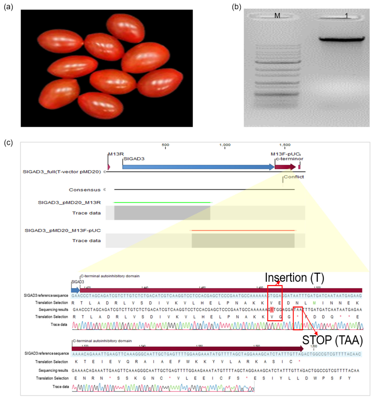
Fig. 1.
Molecular characterization of the target SlGAD3 sequence variant in GABA-enriched tomato. (a) Representative GABA-enriched tomato fruits. (b) Conventional PCR amplification of the full-length SlGAD3 region from GABA- enriched tomato genomic DNA. M, DNA size marker; lane 1, GABA-enriched tomato genomic DNA. The expected amplicon size was 1,925 bp. (c) Sanger sequence analysis of the amplified SlGAD3 region. The PCR product was cloned into the pMD20 T-vector and sequenced bidirectionally using M13R and M13F-pUC sequencing primers. Sequence comparison with the NCBI RefSeq SlGAD3 sequence (GenBank accession No. NM_001246898.2) identified a single-thymine insertion within the region encoding the C-terminal autoinhibitory domain. The insertion altered the downstream reading frame and introduced a premature TAA stop codon. The enlarged region indicates the thymine insertion and the corresponding sequence chromatogram.
Construction and Sequence Verification of the Sequence-Traceable Positive-Control Plasmid
A sequence-traceable positive-control plasmid was constructed for conventional PCR and TaqMan real-time PCR detection of the target SlGAD3 sequence variant. A 120-bp synthetic positive-control insert was cloned into the 2,579-bp pMG-Kan vector, resulting in a recombinant plasmid of 2,699 bp (Fig. 2a). The synthetic insert contained the SlGAD3_var1_F-binding site, two plasmid-specific spacer sequences flanking the GABA-TaqMan probe-binding site, and the SlGAD3_WT_R-binding site. The primer- and probe-binding regions required for routine amplification and fluorescence detection were retained, whereas the two internal spacer sequences were designed to differ from the corresponding GABA-enriched tomato genomic sequence. Sanger sequencing confirmed that the recombinant plasmid contained the intended 120-bp synthetic insert (Fig. 2b). The SlGAD3_var1_F-, GABA-TaqMan probe-, and SlGAD3_WT_R-binding regions were preserved as designed, and both plasmid-specific spacer sequences were correctly incorporated. No unintended nucleotide substitutions, insertions, or deletions were identified within the analyzed insert. Alignment of the plasmid-derived amplicon with the corresponding GABA-enriched tomato genomic sequence showed distinct nucleotide differences within the two spacer regions while retaining the assay-binding regions. These sequence differences enabled plasmid-derived amplicons to be distinguished from the corresponding tomato genomic amplicon by post-amplification sequence analysis. Extended Sanger sequence verification of the constructed positive-control plasmid, including the synthetic target region and adjacent vector sequences, is provided in Supplementary Fig. 2.
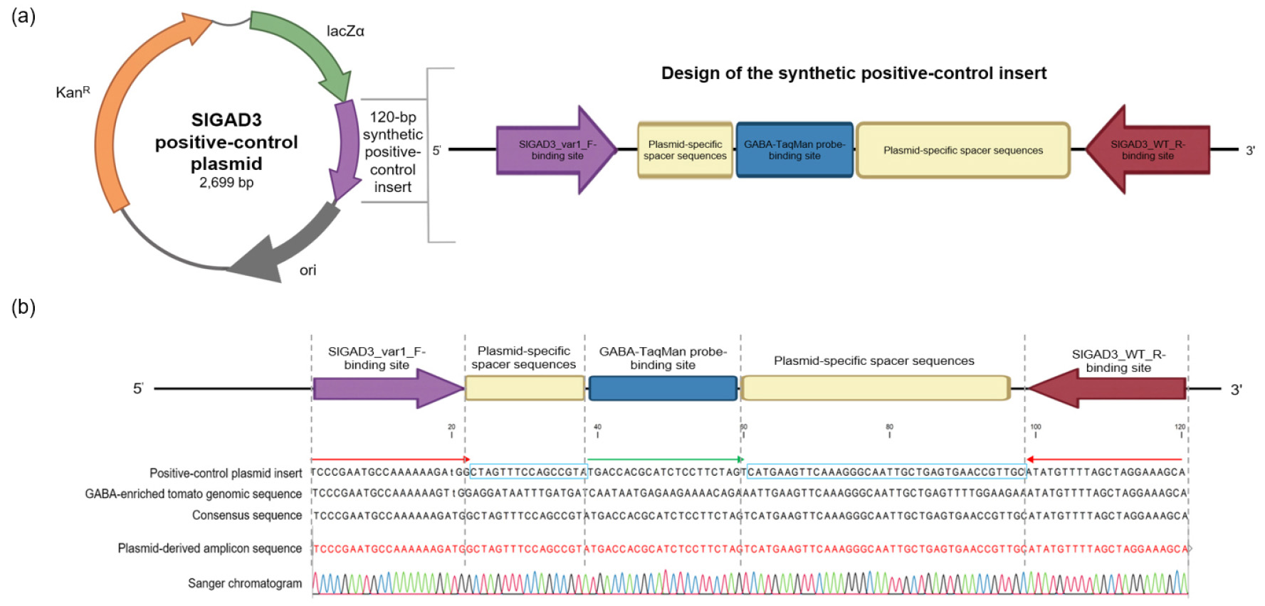
Fig. 2.
Design and sequence verification of the sequence-traceable positive-control plasmid. (a) Schematic representation of the 2,699-bp positive-control plasmid and the 120-bp synthetic insert. The recombinant plasmid consisted of the 2,579-bp pMG-Kan vector backbone and the 120-bp synthetic positive-control insert. The insert contained the SlGAD3_var1_F-binding site, two plasmid-specific spacer sequences flanking the GABA-TaqMan probe-binding site, and the SlGAD3_WT_R-binding site. KanR, kanamycin-resistance gene; lacZα, alpha fragment of the lacZ gene; ori, origin of replication. (b) Sequence alignment and Sanger sequence verification of the 120-bp synthetic positive-control insert. The positive-control plasmid insert was compared with the corresponding GABA-enriched tomato genomic sequence and the plasmid-derived amplicon sequence. The two plasmid-specific spacer regions contained sequence differences from the corresponding tomato genomic sequence, whereas the primer- and probe-binding regions required for amplification and fluorescence detection were retained. The Sanger chromatogram confirmed the intended sequence of the plasmid-derived amplicon. The plasmid-specific spacer sequences serve as post-amplification sequence identifiers for distinguishing plasmid-derived amplicons from the corresponding tomato genomic amplicon.
Conventional PCR Reactivity of the Positive-Control Plasmid
The amplification reactivity of the positive-control plasmid was evaluated by conventional PCR using the SlGAD3_var1_F and SlGAD3_WT_R primer set. A single amplicon of approximately 120 bp, corresponding to the expected size of the synthetic positive-control target, was obtained from the plasmid template, with no prominent non-specific amplification observed (Fig. 3a).
The positive-control plasmid was subsequently analyzed using a 10-fold serial dilution series ranging from 101 to 10-13 ng per reaction. The intensity of the expected 120-bp band decreased progressively with decreasing plasmid concentration. In the representative gel, a faint target-sized band remained visible at 10-8 ng per reaction, whereas no clearly distinguishable amplification product was observed at 10-9 ng per reaction or lower concentrations. Because this experiment was designed to characterize serial-dilution reactivity rather than to establish a validated analytical detection limit, the lowest concentration showing a visible band was not designated as a formal limit of detection.
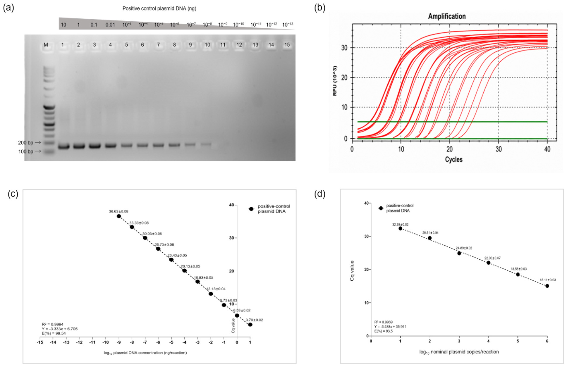
Fig. 3.
PCR amplification characteristics of the sequence-traceable positive-control plasmid. (a) Conventional PCR reactivity of 10-fold serial dilutions of the positive-control plasmid DNA ranging from 101 to 10-13 ng per reaction. M, DNA size marker; lanes 1-15, plasmid DNA concentrations from 101 to 10-13 ng per reaction. The expected amplicon size was 120 bp. (b) Representative TaqMan real-time PCR amplification profiles obtained from serially diluted positive-control plasmid DNA using the SlGAD3_var1_F/SlGAD3_WT_R primer set and the GABA-TaqMan probe. (c) Mass-based relationship between positive-control plasmid concentration and Cq value over the evaluated dilution series. Values are presented as mean Cq ± SD. This analysis was used to characterize concentration-dependent amplification behavior and was not used to assign a formal detection limit. (d) Copy-number-based standard curve generated using six nominal plasmid concentrations ranging from 10¹ to 106 copies per reaction. Values are presented as mean Cq ± SD of technical triplicates. Linear regression yielded y = -3.488x + 35.961, R2 = 0.9969, with an amplification efficiency of 93.5%.
TaqMan Real-Time PCR Amplification of the Positive-Control Plasmid
The positive-control plasmid was evaluated by TaqMan real-time PCR using the SlGAD3_var1_F/SlGAD3_WT_R primer set and the GABA-TaqMan probe. The mass-based serial dilution series generated concentration-dependent amplification curves, with progressively higher Cq values as the amount of input plasmid DNA decreased (Fig. 3b). The corresponding mass-based Cq response showed a strong concentration-dependent relationship over the evaluated dilution series (Fig. 3c). An additional qPCR experiment performed independently on a different day showed a similar concentration-dependent Cq response (Supplementary Fig. 3). The independent run yielded a regression equation of y = -3.328x + 6.750 with an R2 value of 0.9997. These findings support inter-run consistency of the positive-control plasmid amplification under the evaluated conditions.
Copy-Number-Based Standard-Curve Characteristics of the TaqMan Real-Time PCR
Assay a separate copy-number-based standard curve was generated using six nominal positive-control plasmid concentrations ranging from 106 to 101 copies per reaction (Fig. 3d). Mean Cq values were 15.11 ± 0.03, 18.56 ± 0.03, 22.06 ± 0.07, 24.89 ± 0.02, 29.51 ± 0.04, and 32.39 ± 0.02 for 106, 105, 104, 103, 102, and 101 nominal copies per reaction, respectively. Linear regression of the mean Cq values against the log10-transformed nominal plasmid copy numbers generated the following Eq. (2):
The standard curve had a coefficient of determination (R2) of 0.9969 and an amplification efficiency of 93.5%. The 95% confidence interval for the slope was -3.758 to -3.218. These values met the predefined analytical criteria and demonstrated consistent amplification of the positive-control plasmid over the evaluated range of 101-106 nominal copies per reaction.
Specificity of the SlGAD3_var1 Conventional PCR Assay
The specificity of the conventional PCR assay was evaluated using the sequence-traceable positive-control plasmid, GABA-enriched tomato genomic DNA, and genomic DNA from 11 conventional commercial tomato samples (Fig. 4). The positive-control plasmid and GABA-enriched tomato genomic DNA were loaded in lanes 1 and 2, respectively, and the 11 conventional commercial tomato samples were loaded in lanes 3-13. A distinct amplicon of approximately 120 bp was detected from both the positive-control plasmid and the GABA-enriched tomato genomic DNA. In contrast, no target-sized amplification product was detected in any of the 11 conventional commercial tomato samples, and no prominent nonspecific amplification was observed under the PCR conditions used. These results indicated preferential amplification of the target SlGAD3 sequence by the SlGAD3_var1_F/SlGAD3_WT_R primer set within the evaluated sample panel.
The plasmid-derived and GABA-enriched tomato genomic amplicons showed similar electrophoretic sizes and therefore could not be distinguished by agarose gel electrophoresis alone. However, as shown by the sequence comparison in Fig. 2b, the plasmid-derived amplicon contained two plasmid-specific spacer sequences that were absent from the corresponding tomato genomic amplicon. Thus, when positive-control carryover is suspected, post-amplification Sanger sequencing can be used to distinguish a plasmid-derived product from the corresponding GABA-enriched tomato genomic product.
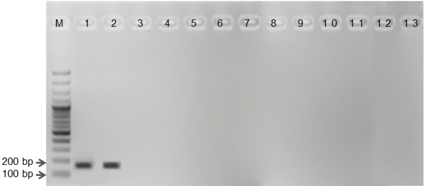
Fig. 4.
Specificity of the SlGAD3_var1_F/SlGAD3_WT_R conventional PCR assay for detection of the target SlGAD3sequence variant. M, DNA size marker; lane 1, sequence-traceable positive-control plasmid; lane 2, GABA-enriched tomato genomic DNA; lanes 3-13, genomic DNA from 11 conventional commercial tomato samples. The expected 120-bp amplicon was detected in the positive-control plasmid and GABA-enriched tomato genomic DNA, whereas no target-sized amplification product was detected in the 11 conventional commercial tomato samples.
Discussion
The present study developed a sequence-traceable positive-control plasmid for conventional PCR and TaqMan real-time PCR detection of the target SlGAD3 sequence variant in GABA-enriched tomato. The construct retained the primer- and probe-binding regions required for the SlGAD3_var1_F/SlGAD3_WT_R assay and incorporated two plasmid-specific spacer sequences within the amplified region. Consequently, the plasmid functioned as an amplification positive control while allowing plasmid-derived amplicons to be distinguished from the corresponding tomato genomic amplicons by post-amplification Sanger sequencing. This combination of routine assay control and internal sequence traceability represents the principal feature of the developed construct. Sequence analysis confirmed a single thymine insertion in the region encoding the C-terminal autoinhibitory domain of SlGAD3. The insertion caused a frameshift and introduced a premature TAA stop codon. Disruption of the C-terminal regulatory region of tomato GAD proteins has previously been associated with increased GAD activity and GABA accumulation. Targeted introduction of premature stop codons into SlGAD2 and SlGAD3 and multiplex editing of genes involved in GABA metabolism have similarly increased GABA concentrations in tomato tissues (Li et al., 2018; Nonaka et al., 2017). Thus, the sequence identified in the present study was consistent with the molecular characteristics previously reported for GABA-enriched tomato and provided the sequence basis for development of the allele-specific assay.
Molecular detection of genome-edited crops can differ from conventional GMO detection because the diagnostic feature may consist of only a single-nucleotide substitution or a short insertion or deletion within an endogenous gene. Such variants do not necessarily contain commonly screened transgenic elements and therefore require prior knowledge of the edited sequence. Editing-site-specific PCR assays developed for genome-edited canola and CAO1-edited rice have demonstrated that short sequence variants can be detected using appropriately designed primers and probes, although discrimination from closely related non-edited alleles remains technically demanding (Chhalliyil et al., 2020; Zhang et al., 2021). The present assay should be interpreted as detecting a defined SlGAD3 sequence variant rather than identifying the genome-editing process by which the variant was generated. Although allele-specific PCR can discriminate the target sequence from closely related non-edited alleles, detection of the short endogenous variant alone cannot demonstrate that it originated specifically from CRISPR/Cas9-mediated editing. An identical sequence could potentially arise through another mutagenic process or occur as a natural sequence variant.
SlGAD3_var1_F was designed according to an allele-specific PCR strategy, with an additional deliberate mismatch introduced near the 3’ terminus to improve discrimination of the target sequence. Deliberate mismatches can enhance allelic discrimination, but excessive destabilization of the primer-template duplex may also reduce amplification of the intended target. SlGAD3_var1_F, which contained one deliberate mismatch, was therefore selected following preliminary evaluation for development of the positive-control assay (Bui and Liu, 2009; Newton et al., 1989; Yaku et al., 2008).
Plasmid-based positive controls provide stable and renewable amplification templates when authentic positive plant material is limited or variable in DNA quality (Caasi et al., 2013; Smith et al., 2006). However, their high target-copy abundance also makes them a potential source of false-positive amplification following accidental carryover. This limitation is particularly problematic when the control and authentic genomic target produce amplicons that cannot be distinguished by routine PCR or fluorescence analysis.
The traceability strategy used in the present study addressed this limitation by incorporating two plasmid-specific spacer sequences while retaining the established primer- and probe-binding regions. A conceptually similar strategy was used in the pBT63 positive-control plasmid, in which a discriminatory restriction site enabled control-derived amplicons to be distinguished from authentic Bt63 products. In the present construct, plasmid-derived and tomato genomic amplicons were similar in size and shared the same probe-binding site; therefore, they could not be differentiated by agarose gel electrophoresis or FAM fluorescence alone. Instead, the two spacer sequences served as post-amplification identifiers that allowed control-derived products to be recognized by Sanger sequencing. Thus, the spacer sequences facilitate investigation of suspected positive-control carryover but do not prevent contamination itself.
The analytical performance of the developed plasmid was considered in relation to previously reported GMO reference plasmids. The pSOY multi-target reference plasmid showed a qualitative PCR limit of detection of 20 copies and a quantitative PCR limit of quantification of 10 copies; its real-time PCR assays exhibited efficiencies greater than 90% and R2 values greater than 0.999 (Pi et al., 2015). The pUC_GM-SB plasmid developed for GM sugar beet H7-1 showed a qualitative PCR detection limit of approximately 10 plasmid copies and a quantitative PCR detection limit of five copies per reaction (Suh et al., 2024). In addition, pUC-RICE5, developed for five GM rice events, supported quantitative detection corresponding to approximately 1-10 copies of rice haploid genomes, with R2 values ranging from 0.993 to 1.000 (Kim et al., 2017). These studies provide useful reference ranges, although they used different target sequences, matrices, validation criteria, and definitions of analytical endpoints.
In the present study, a faint conventional PCR product remained visible at a nominal plasmid concentration of 10-8 ng per reaction. Based on the molecular size of the 2,699-bp recombinant plasmid, this mass corresponds theoretically to approximately 3.4 plasmid copies per reaction. The observed low-concentration reactivity was therefore within the same general order of magnitude as the copy-number ranges reported for pSOY, pUC_GM-SB, and pUC-RICE5 (Kim et al., 2017; Pi et al., 2015; Suh et al., 2024). However, the present value was derived from a representative serial-dilution gel rather than replicate testing near the detection endpoint. It should therefore not be interpreted as a validated LOD or as evidence that the present assay is more sensitive than the previously reported systems.
The copy-number-based standard curve provided a more direct characterization of positive-control plasmid amplification than the mass-based dilution series. Across 101-106 nominal copies per reaction, the assay produced a slope of -3.488, an R2 value of 0.9969, and an amplification efficiency of 93.5%. These values were within commonly accepted ranges for qPCR performance and demonstrated consistent amplification of the synthetic positive-control target across the evaluated copy-number range. In addition, an independently performed mass-based qPCR experiment on a different day produced a concentration-dependent response comparable to that of the primary experiment, supporting inter-run consistency of the assay. However, the independent experiment should not be interpreted as a complete reproducibility assessment because broader between-day, operator, instrument, and interlaboratory variation was not evaluated.
Direct comparison of the present qPCR performance with previously reported reference plasmids should nevertheless be interpreted cautiously because plasmid size, topology, concentration assignment, dilution matrix, primer and probe design, and analytical validation criteria differ among studies. Moreover, the copy numbers used in the present standard curve were nominal values calculated from plasmid DNA mass and molecular length rather than independently assigned absolute copy numbers. Therefore, the copy-number-based standard curve characterizes the amplification behavior of the positive-control plasmid but does not establish its commutability as a quantitative reference material (Bustin et al., 2009; Caprioara-Buda et al., 2012).
Several limitations remain. The plasmid copy numbers used for the copy-number-based standard curve were nominal values calculated from the measured DNA mass concentration and the 2,699-bp plasmid length and were not independently assigned using a reference measurement method such as digital PCR. A formal LOD95, intermediate precision, robustness, long-term storage stability, freeze-thaw stability, and interlaboratory reproducibility were not evaluated. Although an independent qPCR experiment performed on a different day supported inter-run consistency, this limited assessment does not constitute full reproducibility validation. Future work should include independent copy-number assignment, replicate testing around the expected detection endpoint, comparison of circular and linearized plasmids, evaluation of a broader non-target panel, stability assessment, and interlaboratory validation.
Despite these limitations, the developed construct provides a practical sequence-defined amplification positive control for the SlGAD3_var1_F/SlGAD3_WT_R conventional PCR and TaqMan real-time PCR assays. Its principal contribution is the incorporation of two internal plasmid-specific sequence signatures that permit post-amplification identification of control-derived products without modifying the established primer and probe system. The construct may therefore support routine assay verification and facilitate investigation of suspected positive-control carryover. Additional analytical validation, including accurate copy-number assignment, replicate testing near the detection endpoint, assessment of plasmid topology and stability, and interlaboratory evaluation, would be required before the plasmid could be used as a quantitative reference material or assigned a formally validated detection limit.
Conclusion
A sequence-traceable positive-control plasmid was developed for conventional PCR and TaqMan real-time PCR detection of the target SlGAD3 sequence variant in GABA-enriched tomato. The 120-bp synthetic insert retained the primer- and probe-binding regions required for the SlGAD3_var1_F/SlGAD3_WT_R assay and incorporated two plasmid-specific spacer sequences that enabled plasmid-derived amplicons to be distinguished from the corresponding tomato genomic amplicons by post-amplification sequence analysis. The constructed plasmid produced the expected 120-bp amplicon in conventional PCR and showed consistent concentration-dependent amplification in TaqMan real-time PCR. A copy-number-based standard curve over 101-106 nominal copies per reaction yielded an R2 value of 0.9969 and an amplification efficiency of 93.5%, while an independent qPCR experiment performed on a different day showed comparable mass-based amplification behavior. In the specificity assessment, the expected amplicon was detected in the positive-control plasmid and GABA-enriched tomato genomic DNA but not in any of the 11 conventional commercial tomato samples. These findings indicate that the developed plasmid can serve as a sequence-defined qualitative amplification control and facilitate investigation of suspected positive-control carryover through its internal spacer sequences. Further validation, including independent copy-number assignment, formal low-copy detection assessment, stability testing, and interlaboratory evaluation, would be required before use as a quantitative reference material. Importantly, the assay detects the defined SlGAD3 sequence variant but does not by itself establish that the detected variant originated from CRISPR/Cas9-mediated genome editing.